Wellington Eye Centre Optometrist
Have you ever been looking at something and noticed a strange mark floating in front of your eye? You may have even tried to swipe it away, but it doesn’t disappear. This may be what people call an eye floater – and they are very common!
Read on to learn more about pesky eye floaters, what causes them, what can be done about them and how urgently you need to have them checked out.
What are eye floaters?
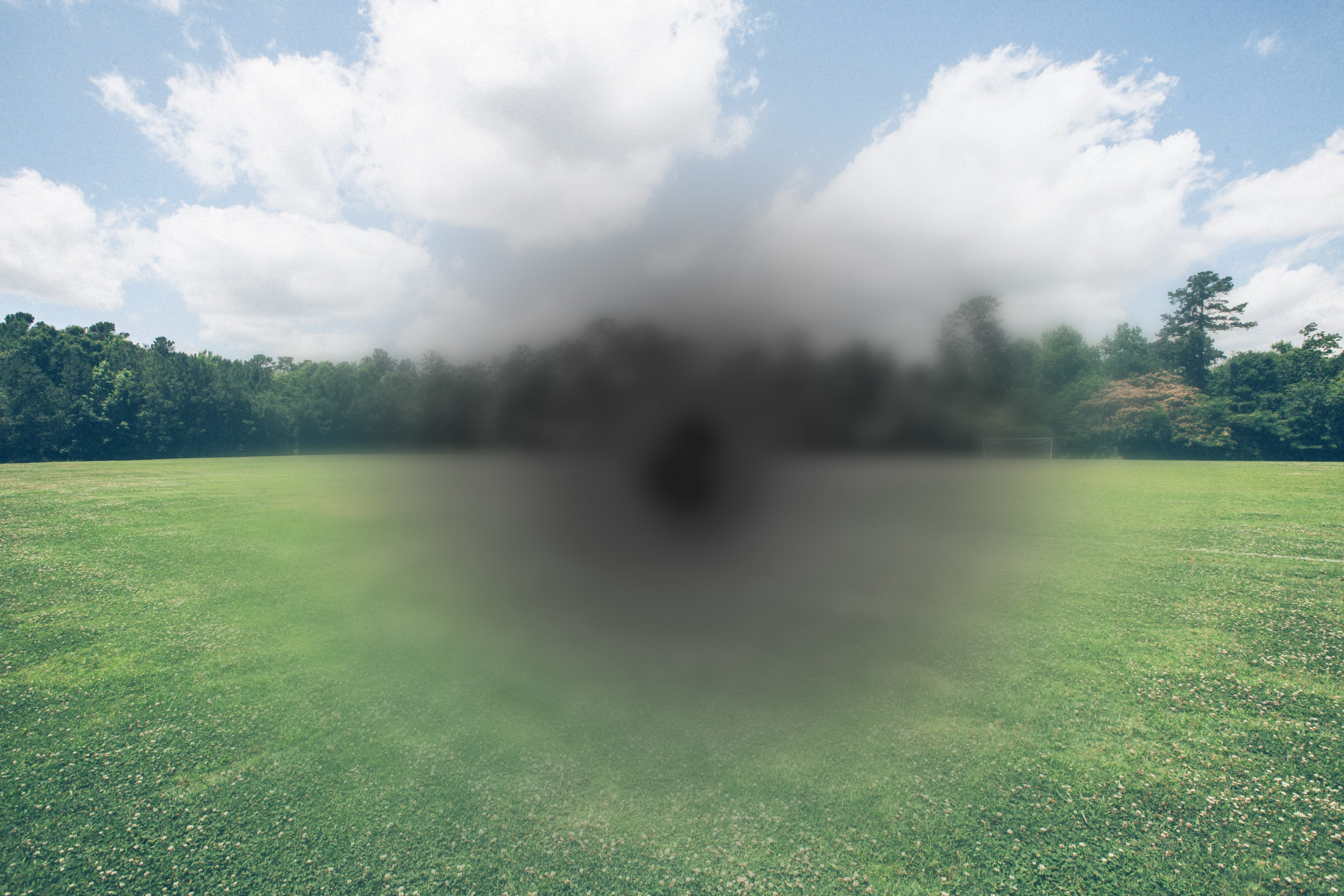
Black spots, threads, wiggly lines or a cobweb like disturbance in the vision is quite common. We call them floaters because they tend to float and drift across your vision with eye movements. The scientific name for them is Muscae Volitantes – the Latin word for “Flying Flies”.
Here’s what eye floaters can look like:
Nearly everyone will report wispy small floaters as they age, but if you have suddenly noticed floaters for the first time, or you notice an increase in the number or size of your floaters, you need to have them checked out that day. We’ll go into that in more detail below!
What causes eye floaters?
Prepare for some medical speak – the cause of eye floaters is all to do with the substances found inside our eyeballs. Here’s how it works:
The eyeball is filled with a jelly like substance called the vitreous. The vitreous is attached to the inside layer of the eye, which is called the retina.
As you age, the vitreous liquefies and shrinks, becoming more mobile. It moves around inside the eye, pulling on the retina, and eventually, breaks away from the retina. This break is called a posterior vitreous detachment (PVD).
A PVD can happen to you with no symptoms whatsoever. Sometimes when it occurs, it releases small bits of debris and old condensed vitreous into the now fluid vitreous. Some people can see these small objects floating in their vision – hence the name eye floaters! They are in fact floating in your fluid vitreous, and what you see is the shadow that these floaters cast on the retina.
When do floaters usually occur?
These floaters can occur at any time in your life. For the most part they’re a sign of the normal ageing eye, and occur during a Posterior Vitreous Detachment. But sometimes, they can be associated with an abnormal change in your eye, called a Retinal Detachment (RD) (more on this below).
You’re most likely to notice your floaters if you’re looking at a plain pale coloured background with a lot of ambient light around you, looking up into the sunny sky, or at a pale office wall.
Here’s a cool video from TEDEd explaining what floaters are:
What can you do to treat eye floaters?
For most people, floaters are an occasional annoyance. For other people, they can be visually debilitating, and in very bad cases an Ophthalmologist can surgically remove them. This surgery, called a vitrectomy, is not performed without thorough consideration. The floaters need to be reducing your vision on the letter chart, and need to be considered constant. In other words, they need to be enough of an issue to outweigh the risks of surgery.
Vitrectomy is a surgical intervention to remove all of your vitreous – the jelly like substance that the floaters are trapped in. It requires general or local anaesthetic and the procedure typically takes one or several hours.
In some cases, eye floaters can be disintegrated with laser intervention. Although, this technique is not commonly used in New Zealand.
Flashes and floaters aren’t associated with laser eye surgery procedures such as SMILE ® , LASIK or PRK. Laser surgery affects your cornea, the front surface of your eye, not your vitreous or retina, which is in a deeper layer of your eye.
When should you see a doctor?
Floaters usually aren’t serious, however you should book an appointment to see your Optometrist or Ophthalmologist if you’re experiencing any of these symptoms:
- Floaters that come on suddenly
- You’re seeing a lot more floaters than you normally do;
- Your floaters are very disruptive to your vision
- Your new floaters have started simultaneously with other new symptoms, such as changes in your vision, grey shadows in your vision, flashes of light, pain and/or severe headaches
- You’ve had retinal detachment in the past
- You’ve had recent eye surgery
- You have other eye conditions affecting your retina
- You’re very shortsighted (myopic) – i.e. your prescription is more than -5.00 diopters
- You have vision in one eye only and experience new symptoms in your good eye
Don’t delay, contact your Optometrist or Ophthalmologist immediately – they will most likely want to see you the same day.
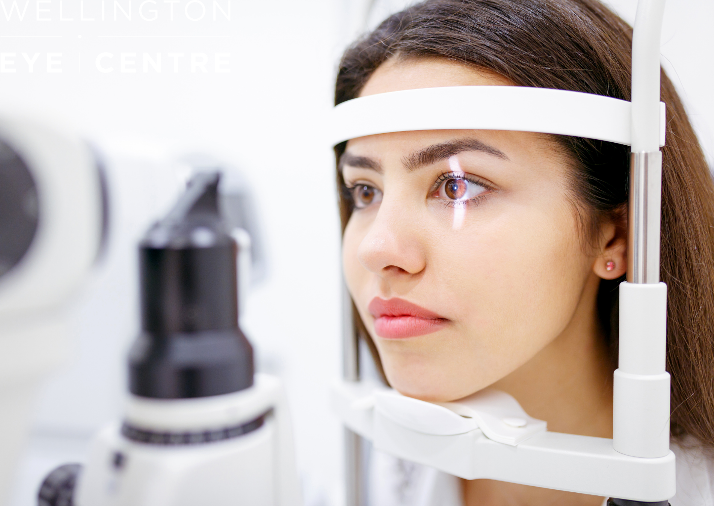
When you visit your Optometrist or Ophthalmologist, they will check your vision on the letter chart, measure the pressure of your eyes, then they will dilate your pupils using an eye drop. Your pupils need to be dilated for the eye care specialist to see the areas of the retina and vitreous that are most likely to be affected in a PVD or RD.
Once your pupil has been dilated, your eye care professional will look inside your eyes to look for signs of retinal and posterior vitreous detachments.
You should note that dilated eyes are more sensitive to light and they have a reduced ability to change focus. So after the eye drops you may have trouble:
- Using a computer or phone screen
- Reading up close
- Driving
- Coping with bright lights (like sunshine, fluorescent lights, or headlights from oncoming cars)
Not everyone is affected in the same way, but being aware of these potential follow on symptoms is important. It shouldn’t last long – the effects of these drops only lasts about 4-6 hours.
What happens if you do have a retinal detachment?
During your examination, if your Optometrist or Ophthalmologist sees a retinal detachment, or enough signs to indicate you’ve had a retinal detachment, they will refer you to a retinal specialist. A retinal specialist can then perform surgery to stabilise the retinal detachment. This surgery is usually done the same day, or the very next morning. Delays in assessing, diagnosing or treating retinal detachments come with a high risk of permanent sight loss. For this reason, your eye care professional will aim to get you assessed and treated as fast as possible.
The surgeries used to resolve retinal detachments vary in complexity. Some are quick, involving a laser to seal the edges of the detachment, which doesn’t usually require an overnight stay at the hospital. Or the eye may require a more invasive surgery, in which case, you may need a general anaesthetic and an overnight stay at the hospital. For this reason, your referring eye care professional may advise you don’t eat, in case you require urgent surgery. So make sure you note when your last meal was.
If treated early enough, retinal detachments have a good prognosis of maintaining your vision. So take any changes in your vision seriously – it’s always better to play on the safe side.
What about flashes of light in my vision?
Before and/or during a PVD or a RD you might also see flashes of light. These flashes could be fine pin pricks of white bright light, an arc, or zig-zag of light. These occur when the retina is being pulled on by the collapsing vitreous, and can occur in one eye or both eyes at the same time.
Flashes may be most noticeable when you’re going from a light to a dark room, in the evenings and often people mention they see them when the lights are out and they roll over in bed.
Flashes are most commonly due to age-related changes in the vitreous and are usually harmless. Sometimes however, flashes can be a pre-warning that the retina is at risk of being torn or detached. Strong pulling on the retina is usually accompanied with increasing and persistent or constant flashes. So, if you’re experiencing those kind of symptoms, make sure you get in touch with your Optometrist or Ophthalmologist as soon as possible.
There are some other serious causes of flashes and floaters:
- Retinal tears
- Infection
- Inflammation
- Hemorrhage
- Head injury
- A significant blow to the head can cause eye flashes and floaters.
Sometimes eye flashes can be caused by a migraine headache. When a migraine is the cause of the flashes of light, they tend to be in both eyes, and last for approximately 20-30 minutes.
Visual disturbances during a migraine can change location in your vision. They can start peripherally and move inwards, or vice versa.
If you develop a headache after these visual disturbances, it’s called a classic migraine with visual aura. Or, if you have the visual disturbances, but no headache afterwards, it’s known as a visual migraine, or an ocular migraine. The causes of both types of migraines are usually the same, and are treated by your GP.
Don’t ignore changes in your vision
Always be proactive and be aware of any changes in your vision!
It’s important to have regular eye checks with your Optometrist. The New Zealand Association of Optometrists recommends that everyone should have an eye examination every 2-3 years. The frequency of your routine examinations may increase to every 6-12month for certain people. Reasons to have more frequent eye examinations could be; family history of eye disease, you’re a contact lens wearer, have diabetes or for younger children with progressing eye sight issues.
Where possible, try and stay with the same Optometrist. Many issues are picked up because the Optometrist notices a change in your eyes. Eye care professionals can detect change much easier if you have baseline records with them.
When you see your Optometrist, tell them of any new symptoms or changes in your vision. Your Optometrist will refer you to an Ophthalmologist if they identify a problem, which requires further investigation or treatment.
How can you check your vision?
Often eye problems can be detected early by identifying any differences between your two eyes. Check your vision one eye at a time. Cover one eye and note what your central and peripheral vision is like; are there any spots, shadows or curtains in your vision? Can you see any flashes or floaters? If there are symptoms, how long do they last for? Then cover the other eye and do the same checks. Are there any differences between the vision in your eyes?
Remember, if you’ve started seeing flashes or floaters contact your Optometrist or Ophthalmologist to book an appointment to have them checked!
SMILE ® is a registered trademark of Carl Zeiss Meditec.

Age Related Macular Degeneration (ARMD)

Everything You Need To Know About Dry Eye

Low-Level Light Therapy for Dry Eyes

Amblyopia and Laser Eye Surgery

Marijuana’s effect on the eyes

Age Related Macular Degeneration (ARMD)

Wellington Eye Centre Sustainability | How We Reduce Waste & Support the Environment

Everything You Need To Know About Dry Eye

What to expect during your Laser Suitability Medical Assessment at Wellington Eye Centre

